Retrosplenial cortex
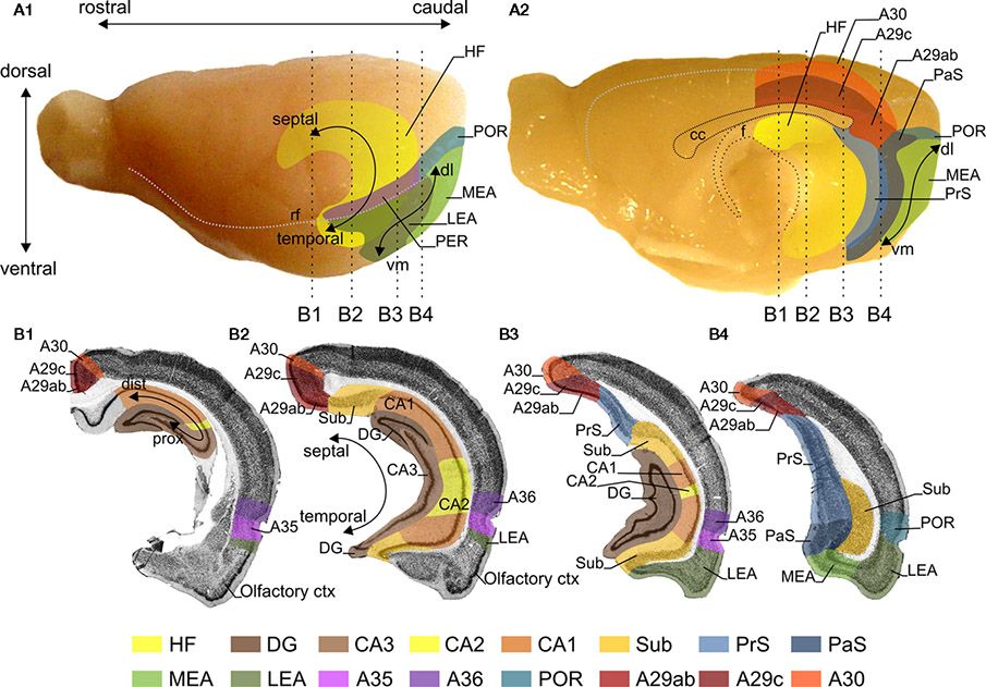
The rat retrosplenial cortex (RSC) is a neocortical structure situated in the midline of the cerebrum. It arches around the dorsocaudal half of the corpus callosum (cc) in the rat, where it is bordered rostrally by the anterior cingulate cortex, caudoventrally by the PHR and laterally by the parietal and visual cortices. The coordinate system that defines position within the RSC is explained in the image below.

Figure 1. Representations of the retrosplenial cortex (RSC), hippocampal formation (HF) and the parahippocampal region (PHR) in the rat brain. Lateral (A1) and midsagittal (A2) views of the rat brain. The RSC is subdivided into A29ab, A29c, and A30. For orientation a rostrocaudal and dorsoventral axis is indicated (A1). The HF consists of the dentate gyrus (DG), CA3, CA2, CA1, and the subiculum (Sub). The PHR is subdivided into the presubiculum (PrS), parasubiculum (PaS), the entorhinal cortex, which has a lateral (LEA) and a medial (MEA) subdivision, the perirhinal cortex (PER; consisting of Brodmann areas (A)35 and A36) and the postrhinal cortex (POR). For orientation in HF the long or septotemporal axis is used [also referred to as the dorsoventral axis; (A1,B2)]. Another commonly used axis is referred to as the proximodistal axis which indicates a position closer to DG or closer to PHR, respectively (B1). The main axis applied in case of PrS and PaS is also the septotemporal axis, in PER and POR a rostrocaudal axis and in the entorhinal cortex is a dorsolateral-to-ventromedial axis [dl and vm; (A1,A2)]. The dashed vertical lines in panels (A1,A2) indicate the levels of four coronal sections (B1–B4), which are shown in (B). The gray stippled line in (A1) represents the position of the rhinal fissure (rf) and in (A2) they represent the global delineation from the dorsal surface of the brain with the midsagittal and occipital surfaces. (B) Four coronal sections of the rat brain; the levels of these sections are indicated in (A). The subfields of the HF, PHR and RSC are color-coded, see color panel below (B). Source: (Sugar et al, 2011).
In the connectome, we follow the nomenclature as described by Vogt in 2004, who subdivides the RSC into four areas referred to as A29a, A29b, A29c and A30. Most of the connectional papers do not separate A29a and b and the combined region we therefore refer to as A29ab. Below follows the summary of the instrinsic connecticity of the RSC and its connectivity with the HF and PHR. These descriptions were published in our publication about the rsc (Sugar et al, 2011) and are an exact copy of that work. The published work contains more detailed descriptions of the rsc connections and we recommend reading this open access publication.
A29a and A29b are distinguished from A29c most strikingly in layer III, which in A29ab has cells arranged in bands parallel to the pial surface, while in A29c layer III is thinner and the pyramidal cell bodies are randomly spaced. An additional way to compare subregions is by looking at chemoarchitectonic features.
A29ab contains parvalbumin-positive cells in layers II, V and VI, which are not as apparent in A29c. In AChE stained sections A29c layer IV shows a widening and increased strength of AChE staining compared to layer IV of A29b.
Cytoarchitectonically, A30 shows an abrupt widening and a less dense packing of layer II/III compared to A29b and A29c. Also, A30 layer IV is wider than in A29b/A29c and A30 layer V neuronal cell bodies tend to be larger. In AChE stained sections, layer I-IV of A30 are evenly and darkly stained, whereas in A29c superficial and deep parts of layer I and layer IV are most densely stained.
Summary of intrinsic retrosplenial cortex connections
There are strong intrinsic connections within the RSC subdivisions. All rostrocaudal levels within both A29c and A30 issue projections to their respective rostrocaudal extents. A29b projections have a strict topography from rostral-to-rostral and from caudal-to-caudal; A29a only has a caudal-to-caudal projection. There are also strong reciprocal connections between the RSC subdivisions. All rostrocaudal levels of one subdivision project to all rostrocaudal levels of all other subdivisions, but there are some exceptions: (1) caudal A29a projects only to caudal A29b, A29c, and A30; (2) caudal A29b does not project to rostral A29c; (3) rostral A29c does not project to caudal A30; (4) rostral and midrostrocaudal A30 only projects to caudal A29b and the return projection from A29b only terminates in caudal A30.
Summary of RSC – HF/PHR Connections
The retrosplenial cortex (RSC) projects to all parahippocampal (PHR) subdivisions and subiculum (Sub). Only the projections of RSC to presubiculum (PrS) and Sub show a topographical organization such that the rostrocaudal axis of origin in RSC correlates to a septotemporal terminal distribution in PrS and Sub. The projections to PrS are among the densest of RSC–PHR connections (Jones and Witter, 2007) and this is particularly true for projections from caudal RSC (Shibata, 1994). A topographical pattern for reciprocal connections has not been identified. Areas 30 and 29c receive input from the whole PHR and Sub, while PrS and Sub are the only areas which project to A29b and A29ab. Dorsal CA1 projects only to A29ab and A29c.
Suggested reading
Sugar, J., Witter, M. P., Van Strien, N. M., & Cappaert, N. L. M. (2011). The retrosplenial cortex: intrinsic connectivity and connections with the (para)hippocampal region in the rat. An interactive connectome Front. Neuroinform, vol 5, 1 - 13 Resolve DOI
References
Jones, B.F. & and Witter, M.P. (2007). Intrinsic connections of the cingulate cortex in the rat suggest the existence of multiple functionally segregated networks Hippocampus 17, 957–976. Resolve DOI
Do you want to read more about the retrosplenial cortex?
We have prepared a pubmed search for you to get started.