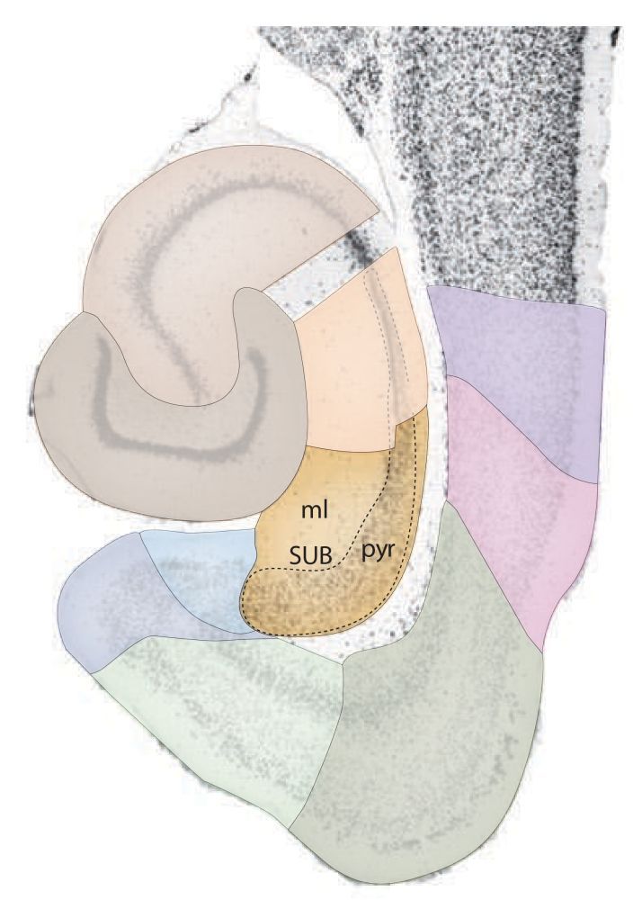
Subiculum
The subiculum is an allocortical structure that stretches between the CA1 field of the cornu ammonis and the presubiculum. It was originally described by Ramón y Cajal (Ramón y Cajal, 1911), but only after it was discovered that the subiculum gives rise to the fornix projections (Swanson and Cowan, 1975), it gained significant interest from the wider scientific community. Sometimes the subiculum is lumped together with structures indicated by the names prosubiculum, presubiculum, parasubiculum and postsubiculum. This conglomerate of structures is called the subicular complex. There is no consensus about the existence of prosubiculum and postsubiculum in the rat. For different species, different arguments are used to falsify or confirm their existence.
In the rat, the prosubiculum may be seen as a oblique transitional region, where CA1 gradually replaces the subiculum. Some argue that the postsubiculum shares the histochemical and connectional characteristics of the presubiculum and therefore prefer to lump it together with presubiculum (e.g. Blackstad, 1956, Honda et al, 2004, Honda et al, 2008), such that the postsubiculum is equal to the septal portion of the presubiculum. Others, emphasize that the postsubiculum has unique histochemical and connectional characteristics (e.g. Rose and Woolsey, 1948, Van Groen and Wyss, 1990) - and that it should considered as a separate region.
The information on this website is written under the assumption that it is better to avoid the terms pro- and postsubiculum and that these regions should be lumped together with the subiculum and presubiculum, in the manner explained above. Apart from the issue with existence of pro- and postsubiculum, the term subicular complex is lumping two categories of cytoarchitectonic structures together. The subiculum resembles allocortex, whereas the pre- and parasubiculum show an increase in the number of layers and are therefore considered to be periallocortex. The pre- and parasubiculum are therefore described on this website as part of the parahippocampal region.
Stratification
In this description of the stratification/lamination of the subiculum, the most superficial layer, close to the pial surface of the brain comes first, followed by the layers below.

- The most superficial layer is referred to as the molecular layer or stratum moleculare and this layer is divided in a superficial and a deep part. The superficial stratum moleculare in the subiculum is a continuation of the molecular layer that runs between CA1 and the presubiculum. The deep stratum moleculare on the other hand, runs as a continuation of the stratum radiatum. (see e.g. Blackstad, 1956)
- The pyramidal cell layer or stratum pyramidale. The border with CA1 is determined by an abrubt wideing of the this layer, abrubt loss of staining in calbindin stained material and increased staining in Timm stained material. The border with the presubiculum is harder to detect, but a noticable decrease in the size of pyramidal cells can be observerd in layers II/III of the presubiculum.
- The stratum oriens, although present in the rest of the cornu ammonus, is not present in the subiculum.
Suggested reading
Boccara, C. N., Kjonigsen, L. J., Hammer, I. M., Bjaalie, J. G., Leergaard, T. B., & Witter, M. P. (2015). . A three-plane architectonic atlas of the rat hippocampal region. Hippocampus, 25(7), 838–857. Subiculum in Hippocampus Atlas
Cappaert, N. L. M., Van Strien, N. M., & Witter, M. P. (2015). Chapter 20 - Hippocampal Formation. In G. Paxinos (Ed.), The Rat Nervous System (Fourth Edition) (pp. 511–573). San Diego: Academic Press.
References
Blackstad, T. W. (1956). Commissural connections of the hippocampal region in the rat, with special reference to their mode of termination. The Journal of Comparative Neurology, 105(3), 417–537. Resolve DOI
Do you want to read more about the subiculum?
We have prepared a pubmed search for you to get started.